Tendons of the Foot JOI Jacksonville Orthopaedic Institute
Common causes of foot pain include plantar fasciitis, bunions, flat feet, heel spurs, mallet toe, metatarsalgia, claw toe, and Morton's neuroma. If your feet hurt, there are effective ways to ease the pain. Some conditions specific to the foot can cause pain, less movement, or instability. Verywell / Alexandra Gordon.
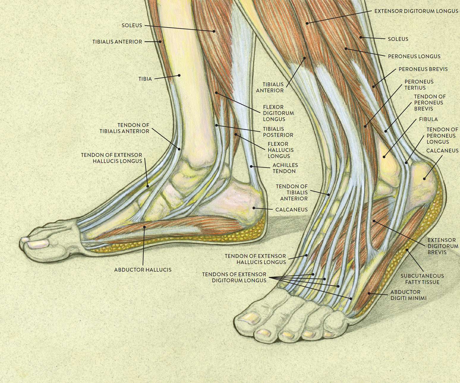
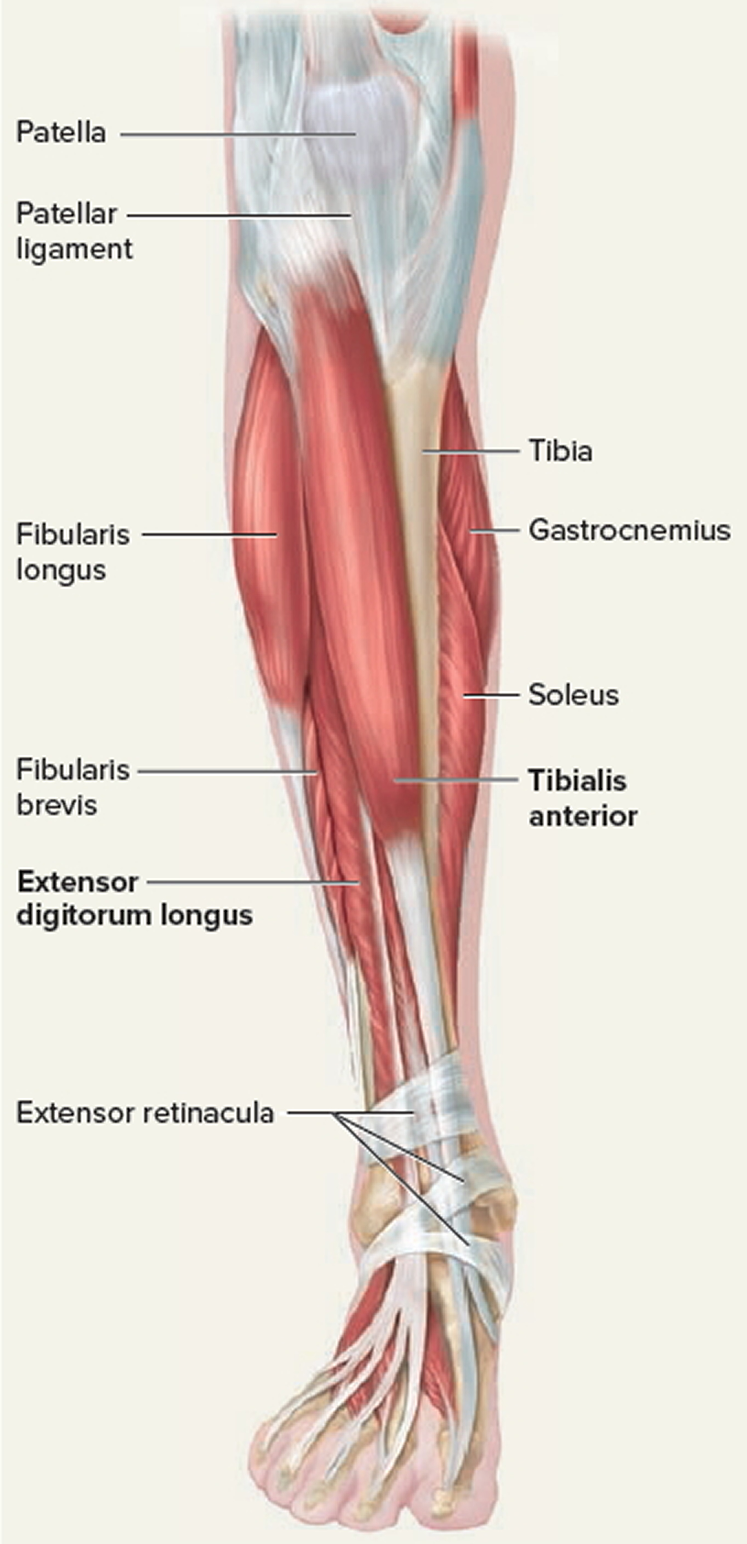
Muscles of the Leg and Foot Classic Human Anatomy in Motion The
Overview What are foot ligaments? Foot ligaments are strong bands of tissue that connect various bones in your foot. The ligaments in your foot help stabilize it. They also provide necessary support to the arches in your feet. Because there are so many bones in the foot, there are also numerous ligaments connecting them.
ligaments and tendons of foot netter CoreWalking
This tendon in the back of the calf and ankle connects the plantaris, calf, and soleus muscles to the heel bone. It stores the elastic energy needed for running, jumping, and other physical.

Loading... Human anatomy chart, Foot anatomy, Nerve anatomy
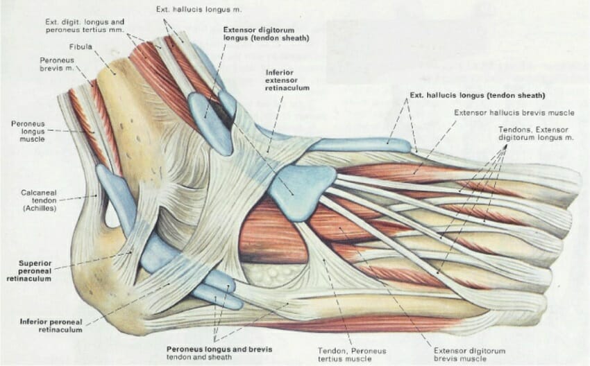
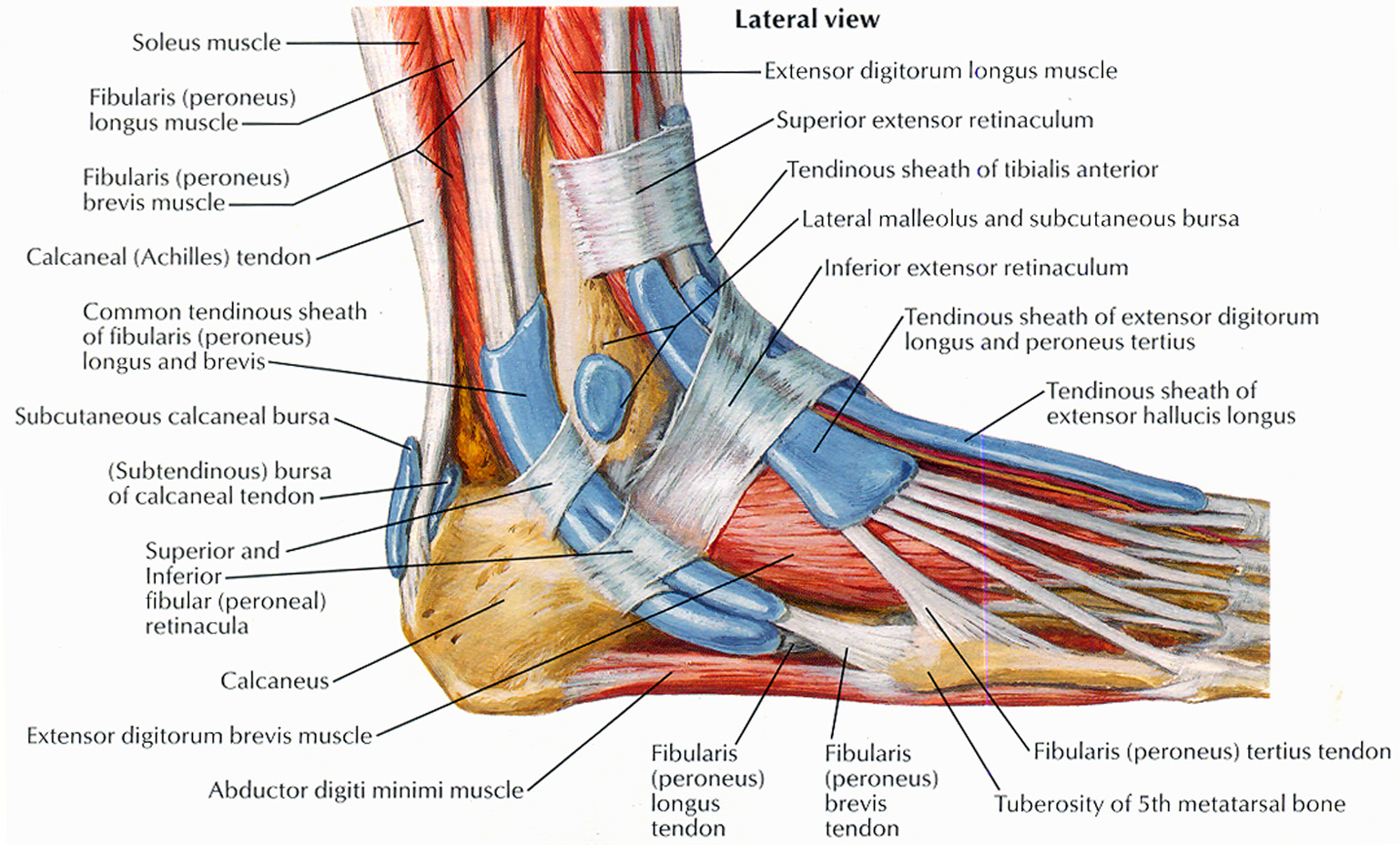
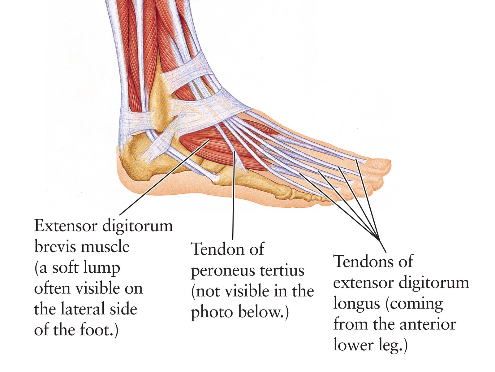
The extensor digitorum brevis is a small, thin muscle which lies underneath the long extensor tendons of the foot. Attachments: Originates from the calcaneus and inferior extensor retinaculum. It attaches onto the long extensor tendons of the medial four toes. Actions: Extension of the lateral four toes. Innervation: Deep fibular nerve.
Tendon Function, Arm, Hand Tendons Leg and Achilles Tendons
1. Foot Bones When thinking about foot and ankle anatomy, we usually divide the into three categories: the hindfoot, midfoot and forefoot. the hindfoot comprises of the ankle joint, found at the bottom of the leg. This is where the ends of the shin bones, the . Underneath this is the heel bone, aka the
Foot (Anatomy) Bones, Ligaments, Muscles, Tendons, Arches and Skin
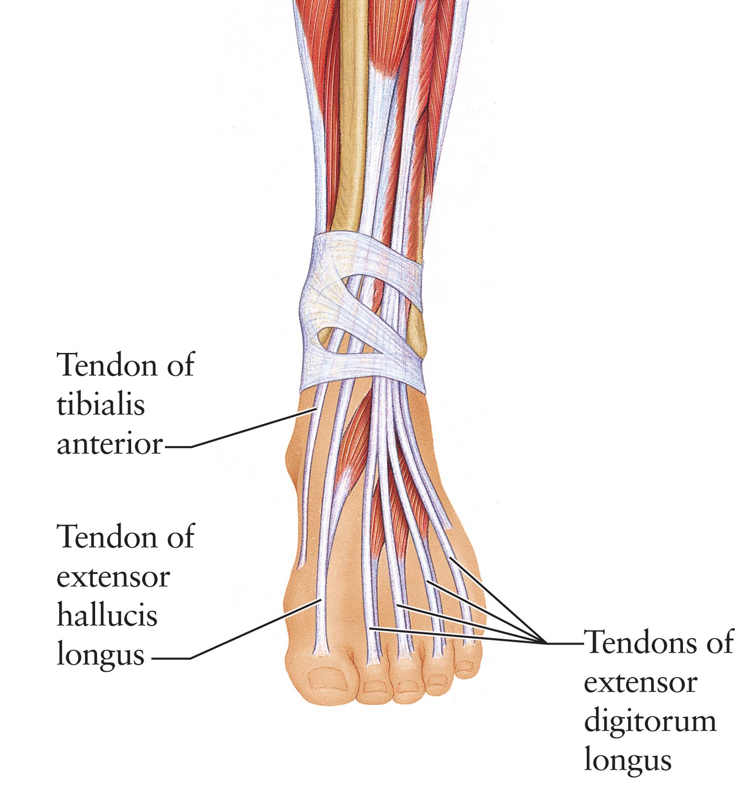
Tendons in the Foot Diagram: Understanding the Anatomy and Function. The foot is a complex structure composed of bones, muscles, ligaments, and tendons. While all these components work together to support our body weight and facilitate movement, it's the tendons in the foot that play a crucial role in transmitting the force generated by the.
Tendinopathy causes, symptoms, diagnosis, treatment and exercises
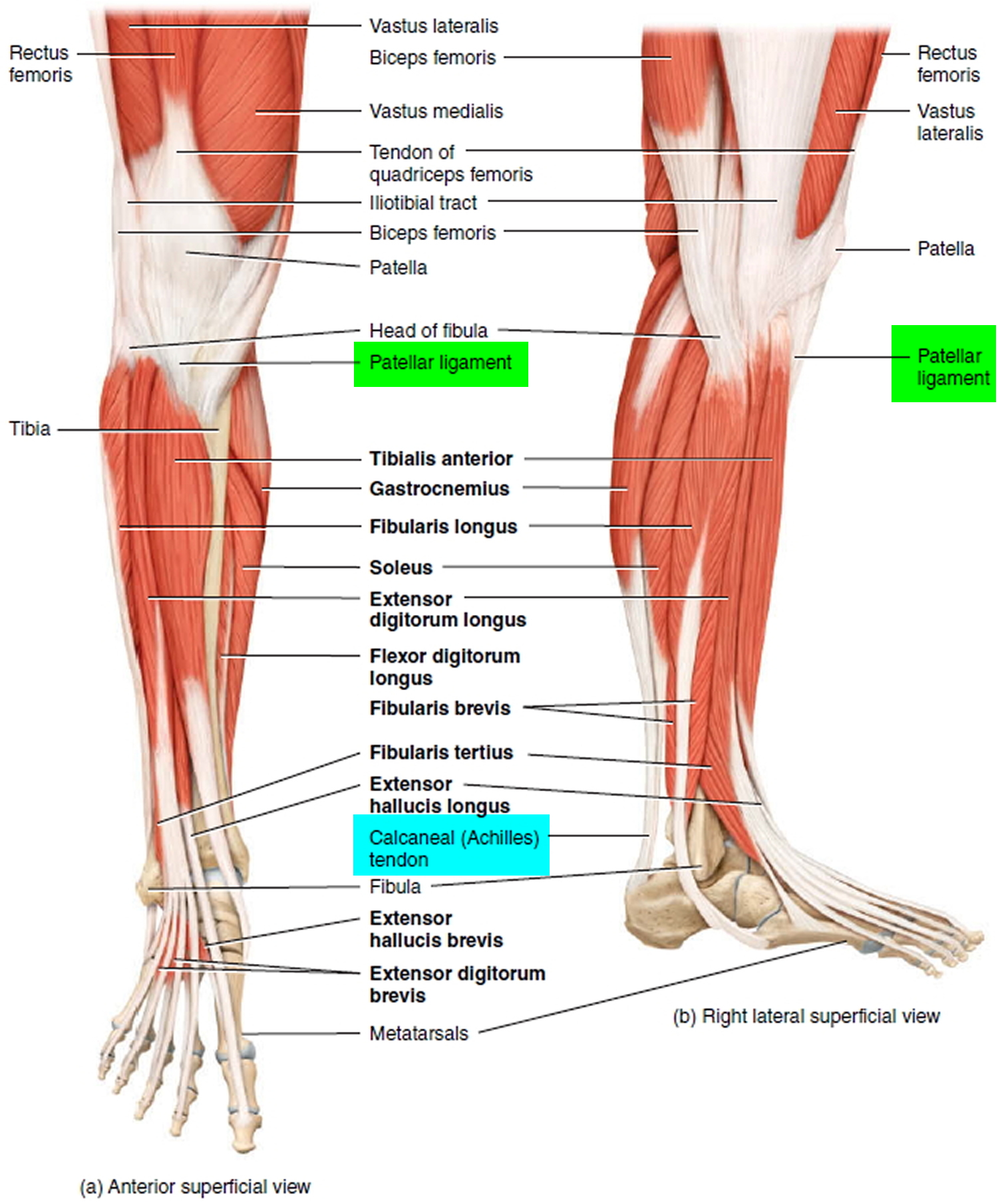
4 Main Motions of the Foot Dorsal flexion (pulling the foot and toes upward): The main tendon for this movement is the Anterior Tibialis. Plantar flexion (pointing the foot and toes downward): The main tendon for this is the Achilles Tendon. Inversion (turning the foot inward): The main tendon for this is Posterior Tibialis Tendon.
Human Anatomy for the Artist The Dorsal Foot How Do I Love Thee? Let
A tendon sheath is a membrane that wraps around a tendon, which allows the tendon to stretch and prevents it from adhering to the overlying fascia. This sheath also produces a fluid, known as synovial fluid, which keeps the tendon moist and lubricated.

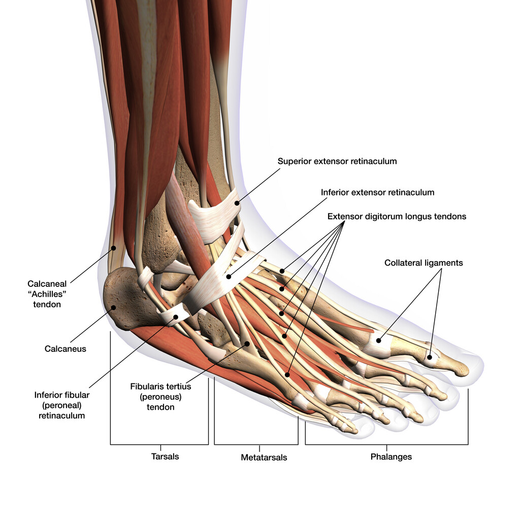
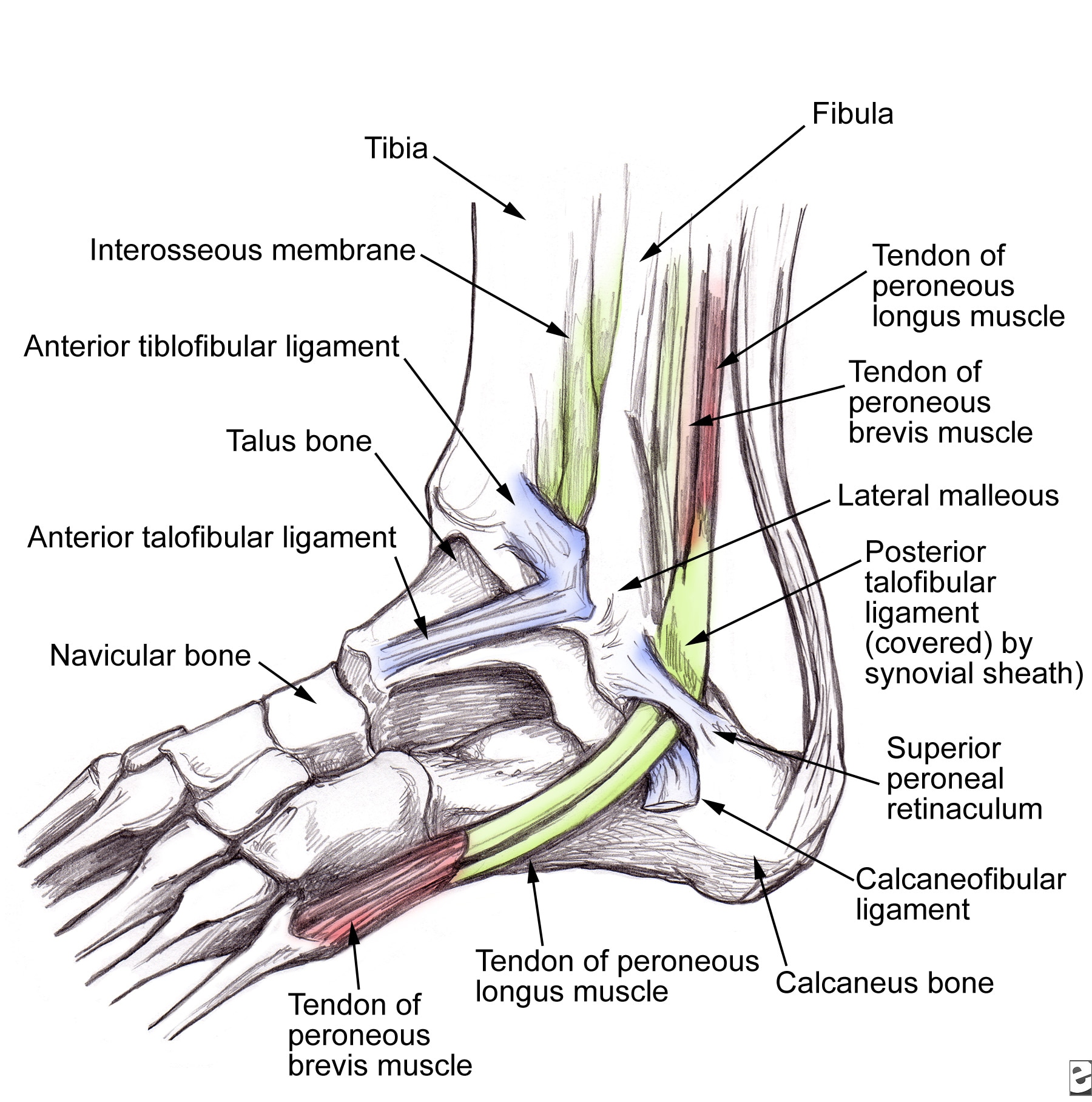
Diagram showing the tendons and ligaments of the ankle and foot
1/2 Synonyms: Talocrural joint The foot is the region of the body distal to the leg that is involved in weight bearing and locomotion. It consists of 28 bones, which can be divided functionally into three groups, referred to as the tarsus, metatarsus and phalanges.
Strained Peroneal Tendon...? run.around.aroo
The foot contains 26 bones, 33 joints, and over 100 tendons, muscles, and ligaments. This may sound like overkill for a flat structure that supports your weight, but you may not realize how much work your foot does! The foot is responsible for balancing the body's weight on two legs - a feat which modern roboticists are still trying to replicate.

Tendon Diagram / Foot Ankle Tendonitis Causes Symptoms Treatment In
Abstract. This chapter discusses the anatomy and pathologies of major tendons of the medial ankle and plantar foot; the posterior tibialis tendon, flexor hallucis longus tendon, and the flexor digitorum longus tendon. The muscles for these tendons originate in the deep posterior compartment of the leg, along with the popliteus muscle.
Foot Anatomy Bones, Muscles, Tendons & Ligaments
The Anatomy of Feet: Bones and Structure Muscles and Tendons in the Foot Ligaments and Joints in the Foot Nerves and Blood Vessels in the Foot Common Foot Conditions and Their Impact on Foot Anatomy Step into the world of feet and explore the intricate anatomy that supports our every step.
Human Anatomy for the Artist The Dorsal Foot How Do I Love Thee? Let
Reviewed By: FPE Medical Review Board Damage to the foot and ankle tendons are a common cause of foot pain, typically caused by overuse, overstretching or an injury. Tendons are thick bands of tissue that connect muscles to bone. When a muscle contracts, the tendon pulls on the bone causing the joint to move.

Foot Description, Drawings, Bones, & Facts Britannica
Foot tendonitis (tendinitis) is inflammation or irritation of a tendon in your foot. Tendons are strong bands of tissue that connect muscles to bones. Overuse usually causes foot tendonitis, but it can also be the result of an injury. Are there different types of foot tendonitis? Your feet contain many tendons.

Tendon Diagram Leg muscles leg tendons hamstrings diagram
A tendon, also known as a sinew, is a fibrous tissue that helps to facilitate this movement. 1 Tendons join muscles to their corresponding bones. Without tendons, your muscles wouldn't be able to make your bones move. Tendons are sometimes confused with ligaments. Ligaments and tendons serve similar purposes, but in different ways.

image lateral_ankle for term side of card Ligament Tear, Ligaments And
It is made up of three joints: upper ankle joint (tibiotarsal), talocalcaneonavicular, and subtalar joints. The last two together are called the lower ankle joint. The upper ankle joint is formed by the inferior surfaces of tibia and fibula, and the superior surface of talus.